Световая микроскопия
Phase-Contrast Equipment
KF·KFA·KFP·KFS·KFZ
PZO · Polskie Zakłady Optyczne · Warszawa - Poland
INTRODUCTION
A normal biological-microscope enables observation of such microobjects only that show some natural contrast to their surroundings, i.e. absorb light in a greater or lesser degree. These are called AMPLITUDE OBJECTS because they change, due to absorption, the amplitude of the light wave. There are, however, microobjects in nature which show mo difference in light absorption as compared with their surroundings, differing only in the refraction index or thickness. Such structures are called phase objects, because they cause only a change in the phase of the light wave that passes through. In a normal microscope, in passing light, they are either totally or almost invisible, because the human eye is not sensitive to changes in the phase of the light wave.
Phase objects have to be dyed, if they are to be seen through a normal biological microscope.
This is troublesome and entails, undesired consequences. Especially live cells and tissues cannot be dyed, because this usually causes their mortification. Thus, the possibilities of observing live microorganisms, that are fully transparent and do not absorb light, by means of a standard microscope system are very limited. A microscope equipped with a phase-contrast device enables observation of phase objects as well as amplitude objects.
Mainly two types of phase-contrast devices are used: positive (Zernike type) and negative.
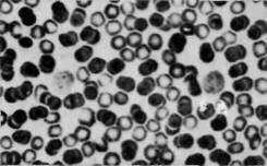
Fig. 1. Corpuscles in Zernike type positive contraste (objective 20X)
The positive phase contrast (Zernike) is more suitable for observation of objects that strongly refract the light, amplitude-phase objects, i.e. objects of an intermediate character between amplitude and phase objects, and is also more suitable for observation of structures with a refraction index smaller than that of the surroundings. Furthermore, in a microscope with positive phase contrast (Zernike type) pictures are obtained that resemble to a high degree those observed in a normal microscope, in a light field — after dyeing of the preparation — which facilitates the identification and differentiation of details.
A negative phase contrast shows generally a greater sensitivity, i.e. enables to detect in the preparation smaller differences in the optical path, provides a greater contrast and a more plastic picture. In a negative phase contrast the background of the picture is dark.
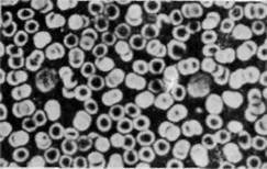
Fig. 2. Corpuscles in negative contrast (phase rings of soot, objective 20X)
Most firms manufacture both positive phase-contrast devices of the Zernike type and negative phase-contrast devices, but this does not yet fully satisfy all needs connected with the observation of objects that have no natural contrast. The Polish Optical Works manufacture besides the positive phase-contrast device KF of the Zernike type and the negative phase-contrast device KFA with phase rings of soot, two new ones: the positive phase-contrast device KFS with dielectric - soot rings, and the phase device with variable contrast KFZ. The KF device has the features of the mentioned positive phase-contrast device of the Zernike type whereas the KFA device shows the features of the a.m. negative phase-contrast. The KFS device has the same advantages and range of application as the KFA type; moreover, it produces a stronger contrast picture which may be desirable for making photographs of microobjects. In view of the positive type of the contrast, pictures seen with the KFS device hove a light background with dark details -similar to pictures observed in light field.
Special attention merits the phase device KFZ with variable contrast. It enables observation both in positive contrast of the Zernike type and in negative contrast KFA, and also provides the possibility of quick and smooth transit from one to the other. Besides these operational advantages the KFZ device produces a stronger contrast picture with better differentiation of details and more sensitive than normal equipment.
DESIGN OF PHASE-CONTRAST EQUIPMENT
The full set of phase-contrast equipment manufactured by the Polish Optical Works comprises the following:
- positive phase device, Zernike type, KF,
- negative phase device, KFA,
- positive phase device, KFS,
- phase device with variable contrast, KFZ.
All these devices may be used in the following types of our microscopes with 160mm tube length: ML4, ML5, ML6, MB30.
The Polish Optical Works manufacture also phase-contrast equipment for microscopes with 170 mm tube length, viz.
— negative phase contrast device KFA10 - for the MB10 microscope,
— negative phase contrast device KFA15 — for the microscope MB15, MB1 and MB2.
Each phase contrast device comprises:
- set of objectives (4 pes)
- condenser
- auxiliary microscope
- key for mounting the condensor.
The objectives of phase contrast devices differ externally in having a stripe of respective colour in the top part of the frame and a symbol added after the numbers that denote the magnification and aperture, as shown in the table hereafter.
E.g. an objective with magnification 10x and aperture 0.24 of the phase device KF has a green stripe on the top part of the frame, and is marked: 10/024 Ph 160/-. The number "160" denotes the mechanical length of the microscope tube. The sign "-" placed after the number "160" indicates that the lens may be used with or without a covering glass. The lenses 20x, 40x, 100x, bear in lieu of the sign "-" the number "0.17" which means that the covering glass must be 0.17 mm thick.
The basic feature of all phase devices, with the exception of the KFZ only, is that in the picture focal plane they have a phase ring that can be seen even with the naked eye after the ocular has been removed from the microscope tube.
The objectives of the KFZ device have two phase rings each.
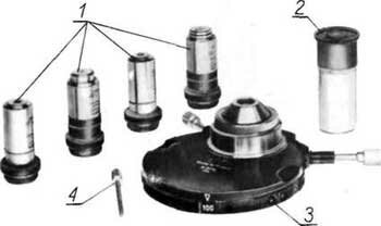
Fig. 3. Main parts of the phase-contrast device: 1. Objectives; 2. Auxiliary microscope; 3. Condensor; 4. Key
All phase contrast devices, except only the KFZ, are equipped with an Abbе condenser K3Ph with an aperture of 1.2. The condenser has an indexing disk with four ring type diaphragms and one free "window". On the periphery of the disk are equidistant marks "0", "10", "20", "40" and "100", and on the fixed frame a pointer is provided for setting the indexing ring. When the "0" mark is in line with the pointer, the free "window" is placed in the path of the rays, and when e.g. the "10" mark is set against the pointer, then the ring diaphragm for the objective 10X is engaged, and so on. The snap lock must snap-in perceptibly to ensure correct positioning of each diaphragm. The condenser hos two centring screws for concentric alignment of the picture of the ring diaphragm with the phase ring in the objective.
Phase device -- Stripe on lens -- Symbol on lens
Positive KF, Zernike type -- green -- Ph
Negative KFA -- yellow -- PhA
Positive KFS -- grey -- PhS
With variable contrast KFZ -- white -- PhZ
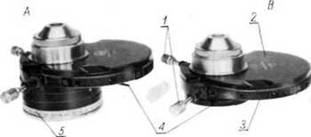
Fig. 4. Condensers of phase contrast devices: A - Condenser K3PhZ, B - Condenser K3Ph
1. Centring screws lor condenser diaphragms; 2. Body; 3. Indexing disc; 4. Lever lor aperture diaphragm; 5. Polarizer
Under the indexing ring the aperture diaphragm is located. The K3PhZ condenser of the KFZ device has four double-ring diaphragms corresponding to two phase rings in each objective and one free "window". Underneath the aperture diaphragm the polarizer is arranged which can be rotated through any desired angle. The polarizer is mounted on slanting guideways and can be removed. When replacing the polarizer it is necessary to make sure that the lock has snapped in.
All phase contrast devices have the same auxiliary microscope MPh. This is used for observation of the phase ring of the objective while the illumination in the phase contrast is being adjusted.
PREPARING THE PHASE CONTRAST DEVICES FOR OPERATION
With the exception of the KFZ type, all phase contrast devices are to be prepared for operation in the same way. Preferably, especially with microscopes with built-in illuminator, the microscope should be set up so as to let the object table and the indexing bowl with the lens seats be positioned directly in front of the observer. The first operation to perform is to screw all objectives into the seats in the indexing bowl. Then the condenser must be mounted in the condenser elevator seat. To do this the elevator should be lowered as far as possible, the condenser introduced from underneath and pushed home so as to locate the condenser centring knobs symmetrically over the shallow recesses in the condenser frame. After pushing home the condenser it is necessary to lock it in position by tightening the grub screw by means of the key provided.
After a suitable ocular (oculars) has been selected and inserted into the tube (tubes) the illumination should be arranged according to the Kohler principle. The respective procedure is as follows.
- Set the condenser indexing disk in position "0" (now the condenser operates like a normal Abbe type condenser).
- Set a 10x or 20x objective over the preparation.
- Connect the illuminator through a transformer to A.C. mains and switch on the light.
- Looking through the ocular bring the preparation picture into focus first by rough and then by fine adjustment.
- Rotating the lever in the rear part of the microscope base reduce the aperture of the field diaphragm.
- Rotating the condenser elevator screw move the condenser so as to bring the picture of the field diaphragm into the plane of the object, i.e. to make it as well as possible visible when the preparation is viewed through the microscope.
- Correct any eccentricity of the field diaphragm picture by means of the condenser centring screws.
- Increase the size of the field diaphragm picture to the dimension of the microscope field of view.
- Reduce the opening of the aperture diaphragm under the condenser indexing disk.
- Looking at the leaves of the aperture diaphragm from underneath, preferably with the aid of a mirror, move the illuminator in a position to obtain on the nearly closed aperture diaphragm a sharp picture of the incandescent lamp filament.
- Remove the ocular from the tube. Increase the picture of the aperture diaphragm, visible against the background of the objective phase ring, a little beyond the outer diameter of the phase ring.
- Replace the ocular in the ocular base tube and use the fine adjustment to bring the picture into focus.
After these preparations the microscope is ready for observation in a "light field". Further details will be found in the operation instructions supplied with each of our microscopes.
To set the illumination for observation in phase contrast adopt the following procedure:
a) Place the condenser indexing disk in position for the respective objective (e.g. using a 20x objective rotate the disk to the position marked "20").
b) Remove the ocular from its tube and replace it by the auxiliary microscope.
c) Move the ocular of the auxiliary microscope until the dark phase of the objective and the somewhat smaller light picture of the condenser ring diaphragm are well in focus.
d) Use the diaphragm centring knob to position the two rings concentrically to each other.
e) Make any necessary corrections, e.g.:
— rotate the aperture diaphragm lever to fit the size of the picture of this diaphragm to that of the condenser ring diaphragm;
— move the illuminator to reproduce the picture of the incandescent lamp filament on the picture of the condenser ring diaphragm;
— rotating the lever in the rear part of the microscope base make the picture of the field diaphragm fit the size of the microscope field of view.
f) Remove the auxiliary microscope from the ocular tube and put in its place the ocular.
For setting the illumination in a phase contrast KFZ perform first all the operations described above under a) through f) with the difference, though, that two pairs of rings have to be positioned concentrically. The inner pair of rings (the aperture ring in the condenser and the phase ring in the objective are for the positive Zernike type contrast, whereas the outer pair is for the negative contrast KFA. Then rotate the polarizer to "extinguish" one of the pairs, leaving the selected contrast and, as in point "f", replace the microscope by the ocular. When using the KFZ device a simple rotary movement of the polarizer suffices to select a positive or negative contrast.
The change-over from one contrast to the other is well visible in the microscope field of view. Moreover, when both contrasts are partly "extinguished", a so-called mixed contrast is produced, suitable especially for the observation of amplitude-phase objects.
The phase device KFZ, besides incorporating two types of contrast — and thereby considerably simplifying operation and making it mcjre convenient — affords the User new possibilities of examining phase objects and amplitude phase objects. When using any phase device it should be borne in mind that any time an objective is changed the setting of the illumination has to be adjusted, the respective ring diaphragm engaged, the openings of the field and aperture diaphragms have to be changed and the concentricity of the aperture and phase rings to be checked — according to the requirements described before. For given conditions and for certain types of examinations and preparations each of the described phase contrast devices may be found to be the most suitable. The correct selecting of the type of phase contrast device is essential for obtaining the desired effect. More detailed information on this subject will be found in the book - "Phase-Contrast and Interference Microscopy" by M. Pluta, edited by PWN (Polish Scientific Editions), 1965.
MAINTENANCE
Phase contrast equipment should be kept in dry rooms, at temperatures from + 55 to 35°C, free of acid vapours and other corrosive substances. During longer breaks in operation the microscope with the phase contrast device should be covered with the canvas or kept in the case.
Use a soft, degreased flannel cloth for cleaning the external surfaces of the metal parts of objective, condenser and auxiliary microscope. In the case of more persevering pollutions, e.g. greasy deposits, finger marks, etc. clean the external surfaces of metal parts with a tampon of clean, degreased cotton-wool soaked in alcohol or ether, wound on a wood stick.
Avoid cleaning external surfaces of optical parts and take care to eliminate conditions promoting pollution of such parts. Should it be necessary to remove dust from the external surfaces of optical parts use a clean beaver hair brush for this purpose. More persevering pollutions of optical surfaces may be cleaned possibly with a clean tampon of degreased cotton wool, soaked in alcohol or ether, and wound on a wood stick. If this does not help, hand over the equipment to a specialized repair shop.
If immersion oil was used, it must be washed off the lens and condenser surfaces with the aid of the solvent (xylol) that is included in the outfit of each microscope manufactured by us.
If the equipment is damaged or the necessity arises to clean the internal surfaces of optical parts or to lubricate any parts, hand over the equipment to a specialized repair shop.
 главная · контакты · книги
 контактная информация, написать сообщение
