Polskie zakłady optyczne (PZO), WARSZAWA
Stereomicroscope MST-131
Description and operation manual
APPLICATION
The stereoscopic Microscope type MST131 gives three-dimensional image of objects and is designed for observation of specimens in the magnification range from 4X - 100X. The microscope can be applied for works which do not require large magnifications, for research work and at making preparations as well as for workshop and laboratory works.
TECHNICAL DATA
Magnification range:
with eyepiece 6.3X - from 4X to 25X
with eyepiece 25X - from 16X to 100X
Field of view:
maximum - 44 mm (at magnification 4X)
minimum - 2 mm (at magnification 100X)
Microscope weight - 6 kg
Weight of the microscope with equipment and case (without transformer) - 10 kg
Case overall dimensions - 380 X 280 X 270 mm
Weight of the stage for observation in transmitted light, with case - 4 kg
Overall dimensions of the stage case - 318 X 240 X 120 mm
Weight of the transformer with the box - 3.5 kg
Box overall dimensions - 320 X 125 X 120 mm
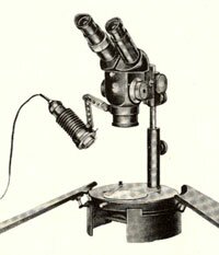
DESIGN
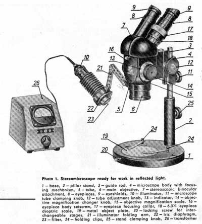
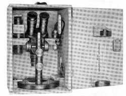
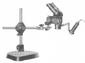
Photo 1. Stereomicroscope ready for work in reflected light.
1 — base, 2 — pillar stand, 3 — guide rod, 4 — microscope body with focusing mechanism, 5 — tube, 6 — main objective, 7 — stereoscopic binocular attachment, 8 — eyepieces, 9 — eyeshields, 10 — illuminator, 11 — microscope tube clamping knob, 12 — tube adjustment knob, 13 — indicator, 14 — objective magnification changer knob, 15 — objective magnification scale, 16 — eyepiece body setscrew, 17—eyepiece focusing collar, 18 —6.3X eyepiece dioptric scale, 19 — metal object plate, 20 — locking screw for interchangeable stages, 21 — illuminator folding arm, 22 — iris diaphragm, 23 —filter, 24— holding clips, 25 — stand clamping knob, 26— transformer
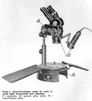
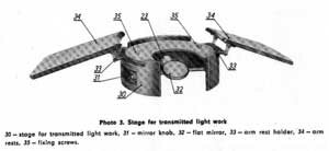
Photo 2. Stereomicroscope ready for work in mixed light (transmitted and refiected)
27 — setscrews, 28 — ground glass plate, 29 — magnification table.
The stereoscopic microscope MST131 being an apparatus of modern design has several special advantages:
- quick change of magnifications which can be effected by turning the magnification changer drum by means of a knob instead of troublesome replacement of objectives applied up to now.
- large and constant working distance amounting approximately to 100 mm creates possibility of a convenient work, specially while making dissections.
- new construction of optical system provides large and uniform field of view up to Ø44 mm.
- inclined stereoscopic binocular body can be applied as required in two opposite directions,
- new optical system applied in the microscope provides natural, perfectly three-dimensional image which is not tiresome for operator's eyes even during long observations.
The microscope is composed of the following fundamental elements: base with stand, tube with main objective, illuminator and magnification changer, inclined stereoscopic binocular body, eyepieces and stage for transmitted light work together with embodied illuminator.
The microscopic slide plate applied for observation in reflected light and the clips holding the preparation are being inserted into the microscope body socket. The plate is painted in white on one side while in black on the other, thus enabling to achieve proper contrast during observation.
The microscope stand of simple and soil id construction assures convenient observation. Releasing the clamping knob located in the stand allows full rotation of the guide-rod together with the body including the tube driving gear without changing sharpness of the image.
The microscope tube is fixed to the guide-rod. Releasing the clamping knob allows the vertical travel of the tube as well as its rotation around the stand. The body with the tube drive is provided with a mechanism which allows smooth shift of the tube being controlled by simultaneous rotation of the L.H. and R.H. knobs in opposite directions.
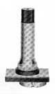
The tube shift range amounts to 46 mm. The main objective with folding arm and illuminator is being mounted in the microscope tube. After having released the set-screw the arm with the illuminator can be rotated around the objective. The folding arm allows the illuminator to be positioned so as to ensure proper illumination of the observed object. The illuminator is provided with a condenser, iris diaphragm and mat blue filter assuring pleasant and uniform illumination of the observed preparation. The iris diaphragm has a maximum opening of 26 mm, minimum opening of 3 mm and is provided with mounting flange ensuring replaceability of photographic filters in holders Ø FF30 manufactured by PZO. A bulb 6V/20W supplied from mains through the transformer TVO-64-6/50. serves as the source of light. The magnification changer drum, by means of which quick change of magnification with constant working distance can be achieved, is embodied in the microscoDe tube. The magnification change is effected by turning a knurled knob with the objective magnification range engraved on it. The value of the magnification is being shown by the indicator. The microscope total magnification can be read from the table.
With telescopes properly set, engagement of a ratchet located inside the body can be felt. The inclined eyepiece body mounted in the tube upper part is being fixed by means of a setscrew.
The eyepiece tubes spacing can be adjusted to suit the pupils distance of observer's eyes. This can be achieved by setting the tubes in their mounts.
The L.H. eyepiece tube is, provided with focusing control collar. Scale engraved on the tube corresponds to the eyepiece 6.3X.
The microscope equipment comprises: the wide field eyepiece 6.3X and 25X and eysshields.
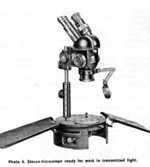
A separate stage on which the microscope is placed is being used for observation in transmitted light. Observed subject is illuminated from below by artifical or day light. A flat mirror mounted in the stage can be adjusted by means of a knob. An illuminator with a bulb 6V/20W supplied from mains through the transformer TVO-64-6/50 provides artificial illumination.
If the examined subject is to be observed simultaneously in transmitted and reflected light, the transformer TVO-64-6/50 produced by PZO allows to connect the both illuminators.
For observation in artificial transmitted light a mat glass plate is being inserted into the microscope base hole, while for observation in transmitted day-light a clear glass plate is being used.
The stage for transmitted light work is provided with hand-rests movable around the stage periphery which ensure easy and comfortable operation, especially during preparation.
The microscope together with its equipment is stored in a wooden case locked with a key.
The stage for transmitted-light work is placed in a separate case locked with latches. The transformer is stored in a cardboard box.
OPTICAL CHARACTERISTICS
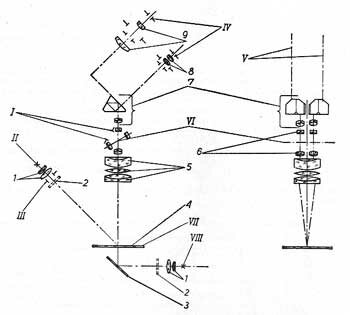
The optical system of the stereomicroscope MST131 is composed of the following main elements: illuminator, main objective, magnification changer, tube lenses, Schmidt's roof prisms and eyepieces.
The upper and lower illuminator are each composed of a bulb 6V/20W and a 2-lens condenser. Colour light filters can be put on the illuminators. The upper illuminator is provided with an iris diaphragm.
The main objective with focal distance 100 mm and diameter 45 mm is a four-lens abjective. As far as its correction is concerned the objective is comparable to modern photographic objectives.
The magnification changer is composed of two pairs of Gallilean telescopes mounted in a rotary drum provided also with an opening. The magnification changer telescopes are corrected specially accurately as they work in both directions. The tube lenses With focal distance 160 mm are also accurately corrected. The Schmidt's roof prisms serve the two purposes:
- to erect the image
- deflection at 45° from the vertical
- to provide changing the interocular distance to suit the observer.
The eyepiece have the large field of view. The microscope is provided with two pairs of eyepieces with magnification 6.3X and 25X.
The optical characteristics of the individual parts of the system are such that:
1) large, 100 mm, working distance is achieved
2) microscope total magnification forms geometric series R5 ace. to PN/N-02100 from 4X to 100X.
The above magnification can fee achieved by prooer setting the magnification changer drum and changing the eyepieces. The microscope total magnification can be obtained by multiplying the objective magnification by the eyepiece magnification. The eyepiece magnifications are engraved on the mounts. The objective magnification is read on the magnification changer scale. Figure 1.6 on the scale corresponds to the objective magnification with the engaged passage opening of the magnification changer drum. The weaker telescope introduces 2.5X factor and in reversed position 1.0X factor, while the stronger telescope introduces 4X and 0.63X factors respectively.
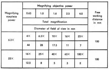
Values corresponding to the telescope are located on the scale opposite to each other. The value 1.6 is being repeated twice.
The magnification series on the changer scale is the following: 0.63X; 1.0X; 1.6X; 4X; 2.5X; 1.6X;
Total magnifications and corresponding diameter of fields of view are given in the Table below:
OPERATION
The stereomicroscope is fixed with a screw to the case bottom for transport. After unscrewing the screw should be kept in its socket in the case.
For work the microscope can be set as required, however the most convenient position of the microscope is with its stand towards the observer.
Put the observed ipreparation on the plate (Photo 1). While using the stage for work in transmitted light insert a mat glass plate (Photo 4) into the microscope base or a clear glass plate for day-light (Photo 5).
The specimen can be illuminated by means of the illuminator mounted on the folding arm (Photo 1) or by means of the illuminator located in the stage (Photo 4) as well as with day-light (Photo 5). The stage illuminator and the upper illuminator can be applied simultaneously giving then mixed illumination. (Photo 2). To obtain an image of the examined specimen:
- release the clamping knob and lift the microscope tube so that distance between the specimen and the main objective amounts to approximately 100 mm,
- set the eyepiece tubes to suit distance between the observer's eye pupils,
- final adjustment of sharpness can be effected by turning the knob and observing the specimen through the microscope,
- adjust the optimal magnification for observation of the given specimen.
The total magnification value can be also selected according to the magnification
Table. Observed object should be properly illuminated.
The illuminator adjustment is being done in the following order:
- after having illuminated the surface of the specimen with upper illuminator, brightness of the obtained light spot should be equalized by means of moving the bulb mount along its axis.
- after having released the setscrew in the ring mounted over the main objective, the whole illuminator can be turned around the main objective and the folding arm with the illuminator is then to be properly positioned and from the correct side thus ensuring optimal illumination of the observed object.
Using the stage for transmitted light work the illuminator light path is to be directed by a fiat mirror. Having reversed the mirror the observation in daylight is possible.
MAINTENANCE
The microscope should be stored in a dry room free of acids or other caustic substances in a temperature from 5°C to 35°C.
If the microscope is not in use for a long period of time, it should be covered or placed in its case. All external surfaces of the optical elements, e.g. main objective, telescopes in the magnification changer drum, inclined body, eyepieces and illuminating system must be cleaned with a special soft flannel rag or beaver brush only. After having completed each test clean the microscope of dust or other impurities taking care not to wipe off the grease from the guides. If the microscope has been damaged or if the inner surfaces of the optical system must be cleaned it should be given either to the proper repair workshop or to the manufacturer.
STANDARD EQUIPMENT
Item Specification Number Symbol Quantity pcs
- Stereoscopic microscope 23420701 1
- Stage for examination 23420702 1
- Eyepiece 6.3X 40750701 2
- Eyepiece 25X 40760701 2
- Inclined stereoscopic binocular body 25090702 1
- Plate 23421002 1
- Mat plate 23420121 1
- Glass plate 23420122 1
- Eyeshield 28010701 2
- Transformer 2897 1
- Bulb 6V/20W 0.9.99.0030 1
- Brush 0.90.7.0100 1
- Flannel rag 0.90.8.0100 1
- Technical description 23502980 1
- Package (set) 23500750 1
- Card of guarantee 23502998 1
OPTIONAL EQUIPMENT (SUPPLIED ON REQUEST)
Item Specification Number Symbol
- Spherical stage 23420706
- Circular rotatable and sliding stage 23420708
- Mechanical stage 23420710
- Long arm stand 4159
- Insects holding support 23420715
- Prism for vertical illumination 23420717
- Adapter for photographic attachment 23490702
- Eyepiece 12X 4071
- Micrometer eyepiece 12X with graticule 4073
- Micrometer eyepiece 12X with scale 4074
- Attachment objective 1,6X 3679
- Drawing attachment 2533
- Polarising device 2781
- Infrastereoscapic accessories 2511
Each of items is delivered in its case
SPHERICAL STAGE
The spherical stage is being set on a metal plate located in the microscope base. The articulated stage plate can be inclined in any direction up to 30° from its horizontal position. The stage surface is covered with rubber plate which protects objects placed on it against sliding down when inclining the stage. Cups with diameters from 45 to 100 mm can be set and fastened on the stage plate by means of movable clips which, after having released the clamping nuts, can be adjusted to suit the required cup diameter. (Photo 9)
CIRCULAR ROTABLE AND SLIDING STAGE
The circular rotatable and sliding stage is being placed into the microscope base socket after having previously removed the reversible metal plate and the holding clips. The stage is immobilized by means of a setscrew (Photo 1, item 20). The table can be rotated by 360° and shifted by 10 mm from its central position in horizontal this making possible comfortable observation and inspection of larger surfaces. The stage surface is provided with a mat glass plate. The stage can be applied for observation at both reflected and transmitted light (Photo 10).
MECHANICAL STAGE
The mechanical stage mounted on an adopter is being placed into the microscope base socket after having previously rerroved the reversible metal plate and the holding clips. The stage is immobilized by means of a setscrew (Photo 1, item 20). The stage can be shifted in longitudinal direction within the range of 30 mm and transverse direction within the range of 65 mm. Longitudinal and transverse shift values can be read on the scales provided with verniers which allow the reading with an accuracy of 0.1 mm. Apart from the cross shifts the stage can be rotated around its axis by 360°. The stage is immobilized by turning a knob located in the adapter.
The stage is provided with an attachment with a mat glass plate and two spare glass plates and with a right-hand holder with spring clip (Photo 11).
LONG ARM STAND
The long arm stqnd is applied for examination of large objects and of complicated shape elements which cannot be observed with help of a standard stand. The long arm stand can be applied either with a round heavy metal base which ensures the microscope stability in every position of arm (Photo 12), or with a bench clamp screwed to the work table (Photo 13). The microscope fixed on the horizontal! arm end can be lifted on the stand up to 480 mm and rotated around the stand axis by 360°. In a horizontal plane the microscope can be shifted together with the horizontal arm up to 360 mm distance. The arm end is provided with a mechanism which permits for microscope additional rotation by 250°. Besides, the horizontal orm together with fixed microscope can be rotated on required level by 360° after having previously secured the stand and released the knob on the cross at the vertical shift.
ATTACHMENT FOR HOLDING INSECTS
For carrying out examination of insects a special attachment has been designed, the insect is being placed in one of the holes provided in the microscope base in place of the holding clips (Photo 14).
The attachment is provided with an articulated cork shaft to which the insects are being pinned. The arm with the cork shaft can be rotated and properly set in desired position which permits for comfortable examination of the object from each side.
POLARISING DEVICE
The polarising device enables the examination in polarised light mainly crystals and anizotropic materials. It is widely used at mineralogy, geology, in various branches of industry and in medicine.
The polarising device consists of an adapter with the polariser, the circular stage with the glass plate, the specimen holder, the analyser and the second polariser, which is put on the illuminator of the microscope base. The holder ensures clamping objects in dimensions from 2 to 6 mm.
PRISM FOR VERTICAL ILLUMINATION
The prism in a special mount, serving for vertical directing of a strong light path from the illuminator to the examined spot, js used for examining holes, slots, smell bores etc. The prism in a mount is being fixed in an objective so that the vertical white line on the mount always coincides with the microscope tube vertical axis (firm trade mark axis) (Photo 16).
The prism is immobilized with a clamping knob. The illuminator should be set by means of a folding arm so that the light path is directed on the prism.
MICROSCOPE ATTACHMENT FOR DRAWING
Microscopic attachment for drawing enables to sketch objects which are observed by the microscope. It permits simultaneous observation of the object and an end of the pencil in the plane of figure. It is very comfortable to attend by the attachment because it doesn't need a blackout room. Leveling the brightness of the object image against the brightness of the background (plane of the drawing) is possible by means of adjustable polaroids Attachment magnification 1X
EYEPIECE 12X
Beside the eyepieces with magnification 6.3X and 25X included into the standard equipment, the eyepieces 12X are additionally supplied on customer's order. These eyepieces permit to obtain additional magnifications of microscope: 8X, 12X, 20X, 32X, and 50X.
MICROMETER EYEPIECES 12X
The micrometer eyepieces 12X permit to make linear measurements of the observed objects by means of the stereoscopic microscope or to determine concentration of elementary particles in the field of view of the microscope.
These eyepieces are provided with a plate with graticule creating an area in dimensions 10X10 mm, divided into 400 elementary fields 0.5X0.5 mm, or with a plate with scale of 10 mm length divided into 100 parts.
ADAPTER FOR PHOTOGRAPHIC ATTACHMENT
The adapter for photographic attachment enables a visual observation and taking photographs in visible light as well as in the infrared. It is a part connecting a stereoscopic attachment with the photographic attachment or an infrastereoscopic attachment with photographic attachment.
INFRASTEREOSCOPIC DEVICE
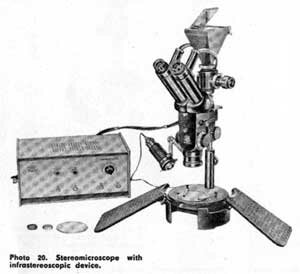
The infrastereoscopic device enables observation of objects in infrared in reflected radiation as well as in transmitted one. It consists of the infra-stereoscopic attachment, a feeder and filters for the work in infrared. Images created by the infrared radiation are changed to visual images by means of a special image transformer mounted in the attachment. The microscope composed of the infrastereoscopic device and stereoscopic microscope, in which instead of stereoscopic attachment with eyepieces is placed infrastereoscopic attachment -thereby becomes an infrastereoscopic microscope for the work in infrared -symbol IM3.
Notice: Drawings and photos contained in the description may differ in some details from actually manufactured products. It is the result of modernisations and improvements introduced continuously by the manufacturer. For eventual differences between description and instrument itself we apologize to our users.
 главная · контакты · книги
 контактная информация, написать сообщение
